
This updated edition provides a comprehensive, detailed approach to veterinary anatomy, offering step-by-step dissection guides for horses and ruminants, with enhanced illustrations and comparative insights.
Veterinary anatomy is the cornerstone of understanding the structure and function of animals, essential for diagnosing and treating diseases. This section introduces key anatomical principles, focusing on horses and ruminants. It emphasizes the importance of anatomical knowledge in clinical practice, surgery, and research. Students and practitioners gain insights into species-specific traits, enabling accurate diagnoses and effective treatments. The guide also highlights the practical skills developed through dissection, such as identifying organs and systems, which are critical for veterinary professionals. This foundational understanding supports lifelong learning and professional competence in veterinary medicine.
Importance of Dissection in Veterinary Education
Dissection is a cornerstone of veterinary education, offering hands-on experience essential for understanding anatomy. It bridges theoretical knowledge and practical application, allowing students to explore complex structures firsthand. By examining tissues and organs, future veterinarians develop critical skills in identification, spatial orientation, and surgical techniques. Dissection fosters a deeper appreciation of species-specific anatomy, enhancing diagnostic accuracy and surgical confidence. It also cultivates problem-solving abilities and attention to detail, which are vital in clinical practice. This experiential learning method remains unparalleled in preparing students for real-world challenges in veterinary medicine, ensuring competence and precision in patient care.
Overview of the 3rd Edition Updates
The third edition introduces enhanced content and modern teaching methods. It features updated anatomical illustrations, detailed dissection steps, and comparative insights between horses and ruminants; New chapters on advanced imaging techniques and their correlation with dissection findings are included. The edition also incorporates feedback from educators and students, improving clarity and organization. Additional digital resources, such as 3D models and video tutorials, supplement traditional learning. These updates ensure the guide remains a leading resource for veterinary anatomy education, blending classic dissection techniques with contemporary tools for a comprehensive learning experience.
Anatomical Overview of the Horse
This section provides a detailed examination of the horse’s external landmarks, skeletal structure, muscular system, and internal organs, forming the foundation for understanding equine anatomy.
External Landmarks and Surface Anatomy
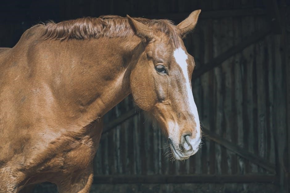
Understanding external landmarks and surface anatomy is crucial in veterinary education. Key points include identifying palpable structures like the withers, spinous processes, and joints. These landmarks guide dissection and clinical procedures. Musculature visibility varies, with areas like the neck and hindquarters revealing significant detail. Surface anatomy also highlights regions for venipuncture and injections, such as the jugular groove. Proper identification ensures safe and accurate interventions. This section provides detailed descriptions and illustrations to aid in recognizing these essential features, connecting anatomical knowledge to practical applications in equine care.
Skeletal and Muscular Systems
The skeletal system of horses and ruminants provides structural support and facilitates movement. Key features include the equine hoof, a specialized adaptation for weight distribution, and the ruminant skull, designed for grinding tough plant material. The muscular system is equally vital, with powerful gluteal and quadriceps muscles enabling locomotion. In horses, the topline muscles are critical for posture and performance, while ruminants rely on abdominal muscles for digestive processes. Understanding these systems is essential for diagnosing musculoskeletal disorders and improving surgical outcomes. Detailed dissection guides highlight anatomical variations between species, emphasizing functional adaptations for movement and digestion.
Cardiovascular and Respiratory Systems
The cardiovascular and respiratory systems in horses and ruminants are intricately designed for efficient oxygen delivery and blood circulation. Horses possess a robust heart and extensive vascular network to meet high-energy demands, while ruminants have a four-chambered stomach influencing respiratory mechanics. Both species exhibit a diaphragm separating thoracic and abdominal cavities. The lungs are divided into lobes, with ruminants typically having more due to their larger body size. Dissection reveals unique adaptations, such as the equine digital cushion and ruminant venous drainage systems. Understanding these systems aids in diagnosing respiratory and cardiac conditions, crucial for veterinary care.
Gastrointestinal and Urinary Systems
The gastrointestinal system in horses and ruminants is specialized for digestion of plant material. Horses have a large cecum for fermentation, while ruminants feature a four-chambered stomach. The urinary system includes kidneys, ureters, and bladder, with variations in structure between species. Dissection highlights differences in renal anatomy and urinary tract design. Understanding these systems is vital for diagnosing digestive and urinary disorders, enabling effective veterinary care and surgical interventions. This section provides detailed insights into anatomical structures and their functional roles in both species.
Anatomical Overview of Ruminants
Ruminants, such as cows and goats, have a four-chambered stomach and specialized digestive system. Their anatomy includes distinct skeletal, muscular, and visceral structures adapted for herbivory and efficient nutrient absorption.
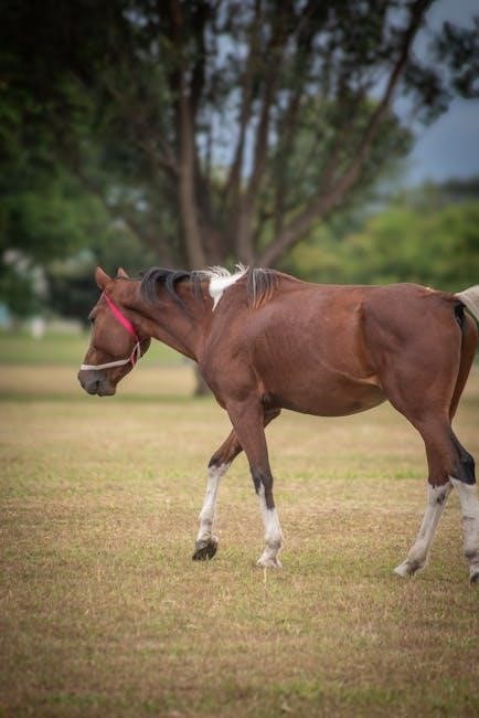
Understanding external landmarks and surface anatomy is crucial for identifying key structures in horses and ruminants. In horses, notable landmarks include the withers, dock of the tail, and fetlock joints. For ruminants, such as cattle, surface anatomy focuses on the loin, brisket, and ruminal areas. These external features guide practitioners in locating internal organs and assessing body condition. Accurate identification of these landmarks is essential for diagnostic procedures, injections, and surgical interventions. This section provides detailed descriptions and visual guides to help students and professionals master the surface anatomy of both species, enhancing their clinical and dissection skills.
The skeletal and muscular systems in horses and ruminants are adapted for unique functional demands. In horses, the skeletal system emphasizes lightweight yet robust structures for speed, while ruminants, like cattle, have a heavier skeleton to support body mass. The muscular system in horses is specialized for powerful locomotion, with notable muscles such as the gluteals and quadriceps. Ruminants exhibit a different muscular arrangement, optimized for grazing and digestion. This section details the anatomical differences, highlighting how these systems contribute to each species’ mobility and productivity, providing a foundation for understanding their functional anatomy and clinical relevance.
The cardiovascular and respiratory systems in horses and ruminants are intricately designed for efficient oxygen delivery and blood circulation. Horses possess a robust cardiovascular system to support high physical demands, with a large heart and extensive vascular network. Their respiratory system is equally specialized, featuring a unique laryngeal prominence to prevent aspiration. Ruminants, such as cattle, have a four-chambered stomach that influences diaphragmatic movement, affecting breathing patterns. Both species exhibit adaptations for their specific environments and metabolic needs, making their cardiovascular and respiratory systems vital for survival and performance, and essential for detailed anatomical study in veterinary education.
The gastrointestinal system of horses is uniquely adapted for digesting plant material, featuring a large cecum and colon for fermentation. Ruminants, such as cattle, have a four-chambered stomach (rumen, reticulum, omasum, abomasum) for breaking down tough vegetation. The urinary system in both species includes kidneys that filter waste and regulate electrolytes, with horses having a more concentrated urine output due to water conservation adaptations. These systems are crucial for nutrient absorption and waste elimination, showcasing evolutionary specializations for their dietary needs and environmental challenges.
Tools and Safety in Dissection
Essential tools include scalpels, forceps, and gloves. Safety measures involve wearing PPE, ensuring proper instrument handling, and maintaining a clean workspace to prevent accidents and contamination.
Essential Dissection Instruments
The primary tools for dissection include scalpels, forceps, and handled knives. Scalpels are used for precise cuts, while forceps assist in tissue handling. Dissection knives with sturdy handles provide control during deeper incisions. Other tools like bone saws and rib cutters are essential for accessing skeletal structures. Adequate instrument maintenance ensures longevity and performance. Proper tool selection and handling are critical for effective dissection, enabling detailed exploration of anatomical structures in both horses and ruminants. These instruments are indispensable for a thorough and safe dissection process, facilitating a deeper understanding of veterinary anatomy.
Personal Protective Equipment (PPE)
Protective gear is essential for safe dissection practices. Gloves prevent skin contact with biological fluids, while lab coats or aprons shield clothing. Goggles or safety glasses protect eyes from splashes, and masks reduce inhalation of particles. Proper hygiene, including handwashing before and after handling specimens, is crucial. Ensuring correct use of PPE minimizes health risks, ensuring a safe and effective dissection environment. These measures are critical for protecting against biohazards and chemicals, promoting a secure learning and working atmosphere during anatomical explorations.
Proper Handling and Maintenance of Tools
Proper handling and maintenance of dissection tools are crucial for safety and longevity. Always use instruments correctly to avoid damage or dulling. Regularly clean and sanitize tools to prevent contamination. Sharpen blades periodically to ensure precision and ease of use. Store tools in a dry, protected environment to prevent rust or damage. Safety measures, such as using cut-resistant gloves, should always be followed. Proper techniques and care extend the lifespan of tools and ensure effective dissection experiences. Regular inspection and maintenance routines are essential for maintaining optimal tool performance and safety in the dissection lab.
Step-by-Step Dissection Guide
This guide provides detailed, sequential instructions for dissecting horses and ruminants, ensuring thorough understanding of anatomical structures through precise, methodical exploration and practical application of dissection techniques.
External Examination and Preparation
Begin with a thorough external examination to identify key anatomical landmarks, ensuring a clear understanding of surface anatomy. Palpate skeletal prominences, such as the withers, spinous processes, and trochanters, to orient yourself. Note the positioning of major vessels and muscle groups. Proper preparation involves shaving and cleaning the specimen to enhance visibility. Ensure the carcass is positioned appropriately for dissection, whether dorsal or ventral recumbency, depending on the procedure. This step is critical for ensuring safety and facilitating an effective dissection process.
Thoracic Cavity Dissection
Begin by making a midline incision from the cervical region to the cranial abdomen, carefully reflecting the skin and musculature to expose the thoracic cavity. Use rib cutters or a saw to remove the sternum and access the thoracic organs. Gently dissect the pericardium to reveal the heart, taking note of its position and major blood vessels. Identify the lungs, trachea, and esophagus, and explore their spatial relationships. Use blunt dissection to avoid damaging delicate structures. This step is crucial for understanding the intricate anatomy of the thoracic organs and their functional interconnections.
Abdominal Cavity Dissection
Begin by making a ventral midline incision from the xiphoid process to the pubic symphysis, carefully reflecting the skin and abdominal musculature. Use a saw to cut through the ribs if necessary, ensuring access to the abdominal cavity. Identify the major organs, such as the stomach, small intestine, large intestine, liver, and kidneys, noting their spatial relationships. Use blunt dissection to explore the mesenteries and ligaments supporting these organs. Pay particular attention to the hepatic and renal blood supply. This step provides critical insight into the abdominal anatomy, essential for understanding digestive and urinary systems in horses and ruminants.

Organ Identification and Exploration
Identify and explore major organs in horses and ruminants, focusing on their anatomical structures, unique features, and comparative differences to enhance understanding of species-specific morphology.
Identifying Major Organs in the Horse
During dissection, identify the horse’s major organs, such as the heart, lungs, liver, and kidneys. Note their anatomical locations and unique features. The heart is a muscular, four-chambered organ located in the thoracic cavity, while the lungs are spongy and situated on either side of the heart. The liver is large and lobed, positioned beneath the diaphragm, and the kidneys are bean-shaped and retroperitoneal. The digestive system includes the stomach, small intestine, and large intestine, with the cecum being a distinctive feature in horses. Use dissection guides to explore these organs and understand their functional roles in the equine body.
Identifying Major Organs in Ruminants
During dissection, ruminants such as cattle exhibit distinct anatomical features. The stomach is divided into four chambers: rumen, reticulum, omasum, and abomasum. The liver is multilobed and located beneath the diaphragm, while the spleen is often darker in color and situated near the stomach. The kidneys are paired, bean-shaped organs in the retroperitoneal space. The heart and lungs are similar to those in horses, with the heart located in the thoracic cavity and lungs expanding in the ribcage. The pancreas is also present, aiding in digestion. Use dissection guides to carefully explore these organs and their unique adaptations for ruminant digestion.
Comparative Anatomy Between Species
Comparative anatomy highlights distinct differences between horses and ruminants. Horses have a single-chambered stomach, while ruminants possess a four-chambered stomach for fermentation. The large intestine in horses is specialized for hindgut fermentation, whereas ruminants rely on their rumen and reticulum. Skeletal structures also vary; horses have a unique cannon bone, while ruminants often exhibit more robust limb anatomy. These adaptations reflect their evolutionary paths and dietary specializations. Understanding these differences is crucial for precise dissection and application in veterinary practice, emphasizing the unique anatomical solutions of each species to their environmental and physiological challenges.
Advanced Dissection Techniques
Mastering advanced techniques involves precise exploration of intricate systems, utilizing specialized tools for delicate structures, and applying anatomical knowledge to complex dissections with accuracy and skill.
Exploring the Nervous System
Dissection of the nervous system requires precision and careful technique to preserve delicate structures. In horses and ruminants, the central nervous system includes the brain and spinal cord, while the peripheral nervous system comprises nerves and ganglia. Use scalpels and fine forceps to gently expose neural pathways, ensuring minimal damage. Identify key landmarks such as the spinal cord segments and cranial nerves. This exploration enhances understanding of neuroanatomy and its functional significance. Proper handling of tissues and tools is essential for accurate identification and documentation of neural structures.
Examining the Lymphatic System
Dissection of the lymphatic system in horses and ruminants involves identifying lymph nodes, lymph vessels, and associated organs like the spleen. Use scalpels and forceps to carefully expose these structures, taking note of their locations near major blood vessels and organs. Pay attention to the mesenteric, inguinal, and cervical lymph nodes, which are key in both species. Proper handling and documentation of these tissues are crucial for understanding their role in immunity and drainage. This section provides detailed guidance on locating and preserving lymphatic structures during dissection, ensuring a thorough comprehension of their anatomical and functional significance.
Understanding the Endocrine System
Dissection of the endocrine system in horses and ruminants focuses on identifying key glands such as the pancreas, thyroid, adrenal, and pituitary. These organs regulate metabolism, growth, and reproductive functions through hormone production. Locate the pancreas near the duodenum and the thyroid glands adjacent to the trachea. The adrenal glands are situated near the kidneys, while the pituitary gland is found at the base of the brain. Carefully expose these structures to avoid damage. Understanding their anatomical relationships and functions is crucial for appreciating their role in overall physiology. This section provides detailed insights into the endocrine system’s organization and significance in both species.

Practical Applications of Dissection
Practical Applications of Dissection involves using anatomical knowledge for diagnostic techniques, surgical procedures, and research, enhancing veterinary medicine’s effectiveness and precision.
Diagnostic Techniques in Veterinary Medicine
Dissection enhances diagnostic accuracy by providing hands-on understanding of anatomy, enabling precise identification of pathological conditions. It aids in refining imaging interpretation and surgical interventions, ensuring effective patient care through practical expertise.
Surgical Applications of Anatomical Knowledge
Anatomical knowledge from dissection is crucial in surgery, enabling precise identification of structures and minimizing complications. It enhances surgical planning, particularly in complex procedures like laparoscopic interventions or orthopedic surgeries. Understanding the spatial relationships of organs and tissues improves technique accuracy, reducing risk to vital structures. Dissection training provides surgeons with the practical skills to navigate anatomy confidently, ensuring safer and more effective interventions. This expertise is vital for both routine and specialized surgical procedures in equine and ruminant care.
Research Contributions from Dissection Studies
Dissection studies have significantly advanced veterinary anatomy research, providing detailed insights into the structural and functional aspects of horse and ruminant anatomy. These studies contribute to understanding anatomical variations, aiding in refining surgical techniques and diagnostic methods. Comparative dissection research highlights species-specific traits, enhancing cross-species anatomical knowledge. Such findings are invaluable for developing new surgical approaches and improving preoperative planning. Additionally, dissection-based research supports the creation of educational resources, fostering better understanding among students and professionals. These contributions underscore the importance of dissection in advancing veterinary science and clinical practice.

The guide underscores the value of dissection in advancing veterinary anatomy, offering a foundation for future research and educational advancements in clinical and surgical practices.
This guide provides a comprehensive overview of dissection techniques for horses and ruminants, emphasizing anatomy, tool safety, and practical applications. It covers external landmarks, skeletal and muscular systems, and organ identification, offering detailed step-by-step dissection processes. The text integrates theoretical knowledge with hands-on practice, highlighting diagnostic and surgical applications. Advanced techniques, such as exploring the nervous and lymphatic systems, are included. The guide also discusses emerging trends in veterinary anatomy, ensuring a well-rounded understanding. By focusing on comparative anatomy and research contributions, it serves as an invaluable resource for students and professionals in veterinary medicine.
Emerging Trends in Veterinary Anatomy
Recent advancements in veterinary anatomy include the use of 3D imaging and virtual dissection tools, enhancing learning and research. Minimally invasive surgical techniques are gaining prominence, reducing recovery times. Wearable technology now monitors animal health in real-time, aiding anatomical studies. Advances in material science are improving prosthetics and implants. Personalized medicine, through biomechanical modeling, tailors treatments to individual anatomy. Telemedicine integrates anatomical knowledge for remote consultations. These innovations are transforming veterinary practice, offering precise diagnostics and treatments while maintaining ethical standards.
Recommended Resources for Further Study
For deeper exploration, utilize online resources like Google Lens for art analysis and conversion tools for quick calculations. Explore advanced search techniques to refine results and access recent studies. Scientific journals from SAGE and the National Academy of Sciences offer valuable insights; The NIH website provides comprehensive guides on laboratory care. Additionally, educational apps like Easy Anatomy offer high-yield notes and 3D atlases. These tools enhance learning and research, aiding in a comprehensive understanding of veterinary anatomy and its practical applications in modern medicine and research.